flow cytometry results for multiple myeloma
We have developed a new statistical diagnostic model that examines the correlation between immunophenotype and clonality as detected by flow cytometry FC and. Many papers were published about this topic - mainly.

Minimal Residual Disease Detection By Flow Cytometry In Multiple Myeloma Why And How Sciencedirect
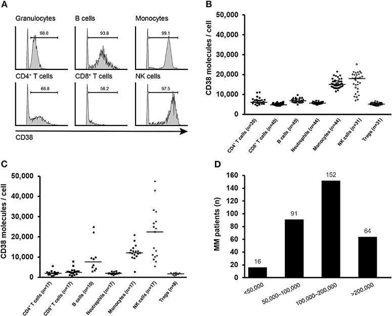
CD38 and CD138 are very common in multiple myeloma and other markers are.

. By ordering this test the clinician acknowledges that additional reflex testing will be performed and billed at a separate. Intervals are Mayo-derived unless. MRD in bone marrow aspirates was assessed using multiparametric next-generation flow cytometry Multiple Myeloma Minimal Residual Disease Panel EuroFlow.
Immunophenotyping is used to determine. Describes reference intervals and additional information for interpretation of test results. May include intervals based on age and sex when appropriate.
We hypothesized that detection of CMCs at. Stringent complete response sCR minimal. If there is a complete.
Apical ligament of dens radiology. Detection of circulating myeloma cells CMCs by flow cytometry in patients with multiple myeloma MM indicates active disease. Cytology and flow cytometry.
Additonal markers will be run if clinically indicated. A combination of Papanicolaou- and Diff-Quik-stained smears is recommended for the evaluation of plasma cells in effusions from patients with multiple myeloma. PURPOSE Assessing measurable residual disease MRD has become standard with many tumors but the clinical meaning of MRD in multiple myeloma MM remains.
Currently multiparameter flow cytometry MFC immunophenotyping is considered to be mandatory for the diagnostic characterization of neoplastic cells and monitoring of. In immunophenotyping flow cytometry identifies protein markers on the surface of myeloma cells. Only you can decsribe the phenotype of plasma cells in MM.
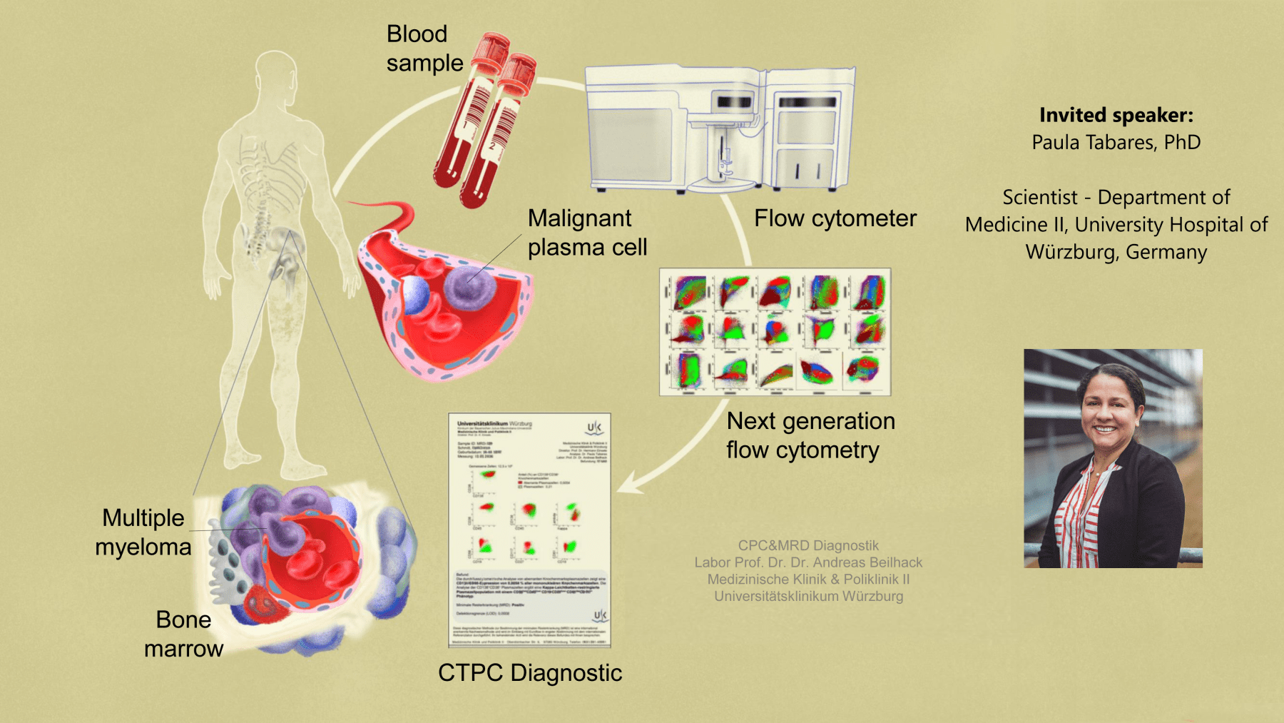
Flow cytometry FC examines individual blood cells using a machine which can separate the cells and detect how light interacts with their surfaces. Multiple myeloma flow cytometry. How dangerous is a 6 cm.
Many myeloma patients have flow cytometry tests performed that show the presence of specific CD markers. Multiple myeloma flow cytometry. First you cannot diagnose multiple myeloma through flow cytometry.
Led zeppelin acoustic guitar lessons. According to results of a study published in the Journal of Clinical Oncology favorable outcomes were observed for patients with newly diagnosed transplant-eligible.

Results Of Newly Diagnosed Transplant Eligible Multiple Myeloma Patients Enrolled In The Forte Trial

Assessment Of Multiple Myeloma Cells And Normal Plasma Cells In Download Scientific Diagram

Minimal Residual Disease In Multiple Myeloma Benefits Of Flow Cytometry Galtseva 2018 International Journal Of Laboratory Hematology Wiley Online Library
Immune Marker Changes And Risk Of Multiple Myeloma A Nested Case Control Study Using Repeated Pre Diagnostic Blood Samples Haematologica

Established And Novel Prognostic Biomarkers In Multiple Myeloma American Society Of Clinical Oncology Educational Book
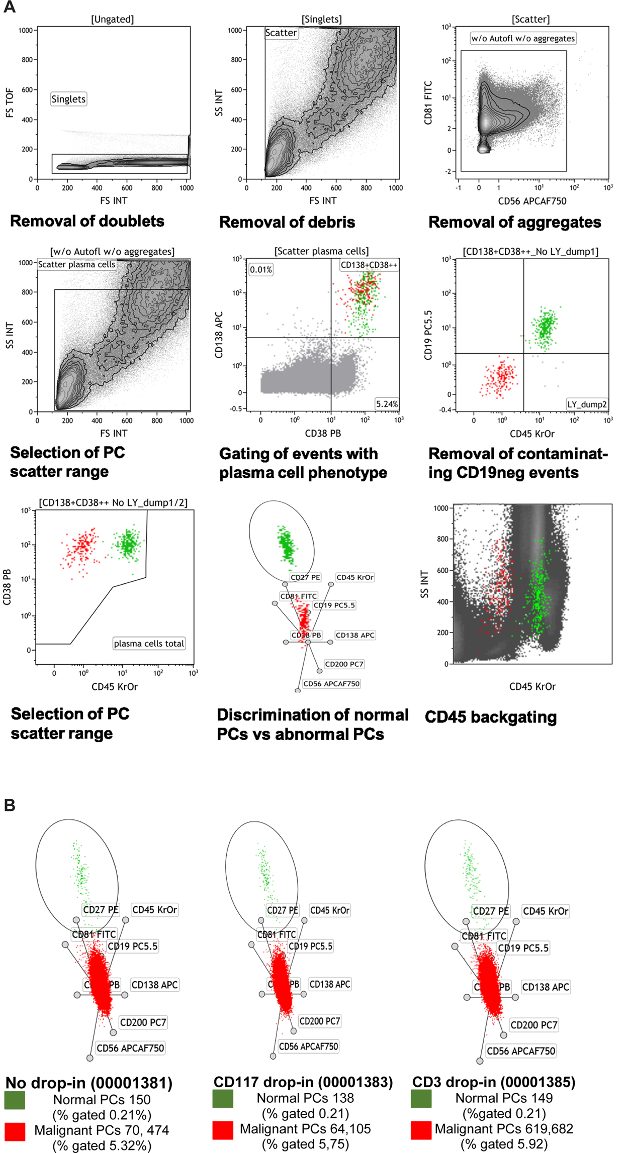
Validated Single Tube Multiparameter Flow Cytometry Approach For The Assessment Of Minimal Residual Disease In Multiple Myeloma Haematologica

Pdf Immunophenotyping By Flow Cytometry In Multiple Myeloma Advantages For Diagnosis And Minimal Residual Disease Monitoring Semantic Scholar

Webinar Monitoring Circulating Plasma Cells In Routine Diagnostics In Multiple Myeloma 2020 10 29 11 00 Am Cet Cytognos S L

Flow Cytometry Characteristic Of Cd4 Cd25 Foxp3 Regulatory T Download Scientific Diagram

Of Flow Cytometry In Plasma Cell Neoplasms Basicmedical Key

Frontiers Isatuximab Acts Through Fc Dependent Independent And Direct Pathways To Kill Multiple Myeloma Cells

Longitudinal Flow Cytometry Identified Minimal Residual Disease Mrd Evolution Patterns For Predicting The Prognosis Of Patients With Transplant Eligible Multiple Myeloma Biology Of Blood And Marrow Transplantation

Immunophenotyping In Multiple Myeloma And Others Monoclonal Gammopathies Intechopen

Flow Cytometric Analysis Of Plasma Cell Myeloma Mature B Lymphocytes Download Scientific Diagram

Flow Cytometer Analysis Is Consistent With Involvement Of Pleural Fluid Download Scientific Diagram

Standardized Assay For Assessment Of Minimal Residual Disease In Blood Bone Marrow And Apheresis From Patients With Plasma Cell Myeloma Scientific Reports

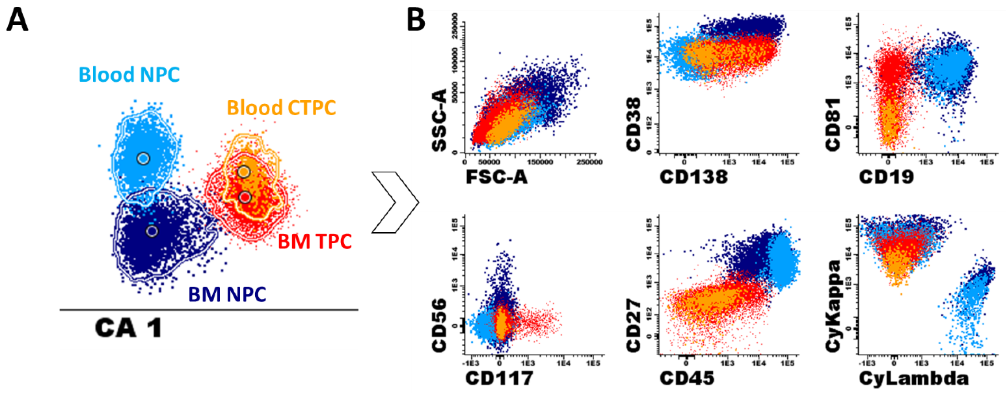
Cancers Free Full Text Detection Of Circulating Tumor Plasma Cells In Monoclonal Gammopathies Methods Pathogenic Role And Clinical Implications Html

Sensitivity Of Flow Cytometry Immunophenotyping Compared With Bone Marrow Morphology In Diagnosis Of Multiple Myeloma Yousof Ea Ahmed Eh Mansor Sg Mohamed Ho Thabet Af Sayed Dm Egypt J Haematol

The Role Of Minimal Residual Disease In Multiple Myeloma Ppt Download